Jump to topic
Search
Instrumentation and Services

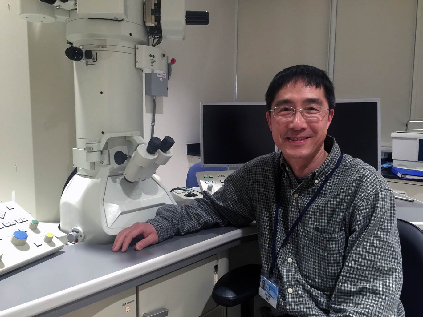
The JEOL 1400 TEM (Room C1727) is capable of generating ultra-structural nanoscale images from fixed cell/tissue samples or multiplexed immune-labeled samples.
- Computer-controlled operations
- Resolution up to 3 Angstroms
- Magnification up to 370,000X
- Capable of collecting data suitable for 3D reconstructions of negative-stained samples
Our mission is to provide high-quality transmission electron microscopy (TEM) services, education and training for the Penn State Health Milton S. Hershey Medical Center and College of Medicine campus, other academic institutions, and private companies at-large.
The lab is equipped with a high-resolution transmission electron microscopes (JEOL 1400) and a full suite of ancillary sample preparation equipment to fit most TEM-related needs. The equipment can provide nanometer-scale analysis for biological, chemical and materials science.
The TEM facility offers multiple levels of service and training including project consultation, education and training for new user self-service and full-service TEM. Available services include sample preparation, fixation, embedding, ultra-microtomy, staining, TEM imaging, immuno-localization (i.e. immunogold EM) and negative staining.
Procedures, Protocols and Forms
Prospective users of the TEM Core can inquire about future projects, either by email, phone or in person. Prospective clients can explore the TEM Core’s fee schedule and set up a time to discuss the project in person with the TEM Core before providing specimens. Investigators should come to the meeting prepared with pertinent information relating to the specific project. At that time a request for specimen submission, including an estimate of costs for the project can be completed so clients and the TEM Core personnel are thoroughly familiar with the proposed project.
The TEM core’s primary goal is to generate high-quality data using electron microscopy. Specimen quality is essential to this process, as without properly fixed and prepared specimens, it is impossible to generate high quality interpretable images. It is our standard policy to review all protocols for incoming specimens and the we reserve the right to reject specimens and/or suggest necessary changes to aid in obtaining adequate and quality results.
Food and beverages are not permitted in the TEM core.
Users of the TEM and all major equipment must demonstrate competency to the TEM Core staff on the instrument before they may use it unassisted, even those who have qualified on similar instruments elsewhere before making the reservation. Users must be able to assess the working condition of an instrument. If an instrument is not working properly, the user should report this to the TEM core staff immediately.
Please register and make the reservation by using the iLab system.
Please follow the instructions at the TEM core.
All publications, press releases or other documents that result from the utilization of any Penn State College of Medicine Institutional Research Resources including funding, tools, servies or support are required to credit the core facility and associated RRID for each core used. Failure to complex may limit your future access to these facilities. Use of an instruments in the TEM core should include the following:
“The TEM Core (RRID:SCR_021200) services and instruments used in this project were funded, in part, by the Pennsylvania State University College of Medicine via the Office of the Vice Dean of Research and Graduate Students and the Pennsylvania Department of Health using Tobacco Settlement Funds (CURE). The content is solely the responsibility of the authors and does not necessarily represent the official views of the University or College of Medicine. The Pennsylvania Department of Health specifically disclaims responsibility for any analyses, interpretations or conclusions.”
Any projects where the TEM core staff prepare the protocol, samples, grids or do the analysis of resulting images are considered to be collaborations. Therefore, the staff should be listed as co-authors. For data collection-only services, on user-provided ready-to-image TEM grids, if the TEM core staff enabled customization of approaches or made significant intellectual input, or overcame extra difficulties to make the work possible, they should be listed as co-authors.
After the paper is published, please send a link to the paper to hchen3@pennstatehealth.psu.edu.
Please refer to the protocols at the TEM Core.
Work With TEM Imaging
Because of limited storage space, we may not store user samples indefinitely. All samples, including blocks and grids, may be returned to the user upon completion of a project.
We strongly recommend a positive and negative control for all experiments. The more information you provide, the easier it will be for us to evaluate and troubleshoot any problems that may arise in the processing of your samples.
Samples that are potentially dangerous to the users or staff of the TEM core may not be brought into the TEM core unless they are rendered harmless according to an approved protocol. The evidence of approval must be on file in the TEM core office. This policy includes but is not limited to bacterial, viral, carcinogenic, and radioactive samples.
The procedures for rendering them harmless must have been approved by the Institutional Biosafety Committee, Research Involving Infectious Agents Committee, and/or the Radiological Safety Board. The evidence of approval must be on file in the TEM core office.
Fixing the samples before bringing them into the TEM core is not sufficient; the full procedure and precautions for preparing the sample for TEM must be approved in writing after complete discussions with the TEM core staff.
Please contact the TEM core.
Services within the TEM cores are managed by the iLab core facility management program. New users must register in iLab to access instruments and services. Follow the quick links below for iLab registration and instructions. Please contact the TEM core to inquire about our services.
Please register and make the reservation by using the iLab system.
