Learn more about the services provided by the Molecular and Histopathology Core.
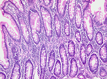
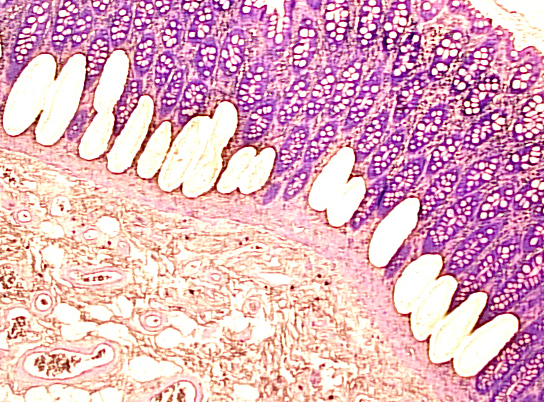
Routine Histology – Processing, Embedding, Sectioning, and Hematoxylin and Eosin Staining
- Fixation and processing of human and animal tissues, as well as cultured cells and tissues Paraffin embedding and sectioning of tissues
- Embedding and cryostat sectioning of fresh, frozen tissues
- Routine H&E staining of human and animal tissues, as well as cultured cells and tissues
- H&E staining for paraffin or frozen sections
Histochemical Stains
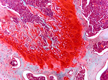
- Histochemical staining of paraffin or frozen tissue sections
- PAS
- Masson Trichrome
- Toluidine Blue
- Gomori’s Aldehyde Fuchsin
- Safranin O
- Verhoeff Elastic
- Cresyl Violet
- Ability to develop and optimize histochemical stains for any tissue type or staining need
Immunohistochemical Stains
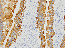
- Immunohistochemical staining oh Human and animal tissues, as well as cultured cells and tissues
- Ability to validate and develop protocols for any antibody
- Mouse-on-mouse/rat immunohistochemistry
- Chromogenic and fluorescent detection methods
- Dual and triple IHC techniques
- All IHC performed on an automated Ventana Discovery XT stainer
In situ Hybridization
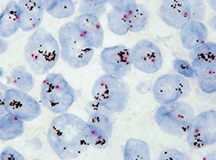
- Chromogenic or fluorescent in situ hybridization protocols
- Ability to develop and optimize protocols for your specific probes
- All hybridizations protocols performed on an automated Ventana Discovery XT stainer
Tissue Microarray
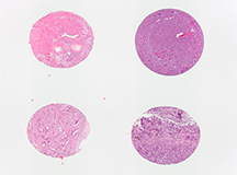
- Paraffin tissue microarrays (TMAs) can be created with any combination of human tissue, animal tissues
or cell culture pellets - Custom-sized and configured arrays are constructed using the Beecher MTA-I
- Tissue core sizes can range from 0.6 mm to 2.0 mm, providing from 40 to over 200 sample cores in one
paraffin block - Microarray slides can be stained using a variety of histochemical or immunohistochemical reactions
Laser Capture Microdissection
- Zeiss PALM Microbeam LCM
- Capable of isolating cells, tissues, or stromal matrix from either frozen and paraffin sections
- Ideal method to insure the purity of the starting material for downstream proteomic and genomic applications
- Capable of operating on an RNAse free level to insure high quality samples for genomic studies
Archival FFPE Human Tissue Samples
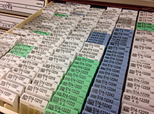
- Archival collection consisting of over 40 years of surgical pathology FFPE specimens
- Searchable database enables the investigator to locate specific patient, tissue or tumor types
- Ability to collaborate with anatomic pathologists to assist in the identification and selection of study samples along with the interpretation and scoring of results
- Perfect for use as control tissues, study samples or TMA sources
- Capable of isolating DNA or RNA from samples
Contact Us
For all technical and project-related questions about the Molecular and Histopathology Core, please contact Marianne Klinger, Research Technician, at mklinger@pennstatehealth.psu.edu or 717-531-1044.
For lab management questions, please contact David Degraff, PhD, Director, at ddegraff@pennstatehealth.psu.edu.